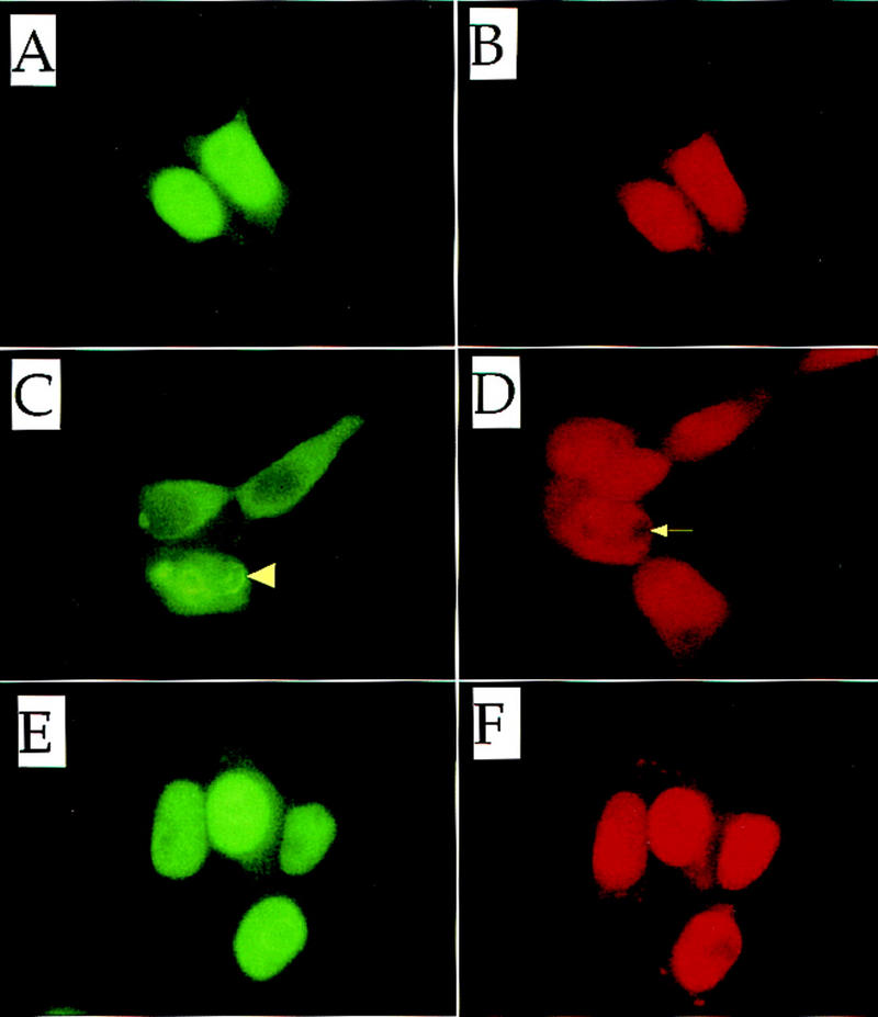
Figure 10.

Subcellular localization of Nrf2. Nrf2 alone (A,B) or both Nrf2 and Keap1 (C,D) were force expressed in 293T cells. Nrf2 and Keap1 were also force-expressed in the presence of 100 μm DEM (E,F). (A,C,E) Localization of Nrf2 was detected using an anti-human Nrf2 antibody; (B,D,F) the same fields stained with propidium iodide. The arrowhead and arrow in C and D, respectively, show the characteristic perinuclear ring structure described in the text.
