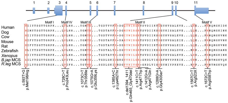
Figure 1.
Alignment of the motif regions in ACSF3 orthologues and the malonyl-CoA synthase enzymes in bacteria. The sequences (see methods) were aligned using MegAlign via the Clustal W method. An additional three amino acids amino-terminal to Motif I are shown. Motif II was aligned independent of the full-length protein to improve the alignment of the ACSF3 and MCS proteins. The ACSF3 alterations identified in the eight subjects and affected dog are indicated. The asterisk (*) indicates the dog variant p.Gly430Ser, which is orthologous to position p.Gly480 in human ACSF3.

