Fig. 1.
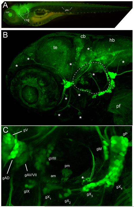
GFP expression of SKIV2L2 enhancer trap line. (A) Whole mount epifluorescence image of Tg(SKIV2L2:gfp) at 4 days- note clusters of GFP+ cells surrounding otic vesicle (dotted line). (B) Confocal microscopy of GFP expression in the peripheral ganglia in the head at 4 days. Asterisks represent neuromasts. te, telencephalon, cb, cerebellum, hb, hindbrain. (C) A detailed view of the sensory ganglia labeled by the SKIV2L2 enhancer. am, anterior macula; gAD, dorsal anterior lateral line ganglia; gAV, ventral anterior lateral line ganglia; gV, trigeminal ganglia; gVII, facial ganglia; gVIII, statoacoustic ganglia; gIX, glossopharyngeal ganglia, gX1-4, vagal ganglia, gM, medial lateral line ganglia, gP, posterior lateral lined ganglia; plln, posterior lateral line; pm, posterior macula; * neuromast.

