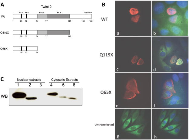
Figure 1.
Subcellular immunolocalization of TWIST2 and its Q119X mutant form reveal nuclear staining. A. Diagram of the TWIST2 protein and mutant forms described for Setleis Syndrome. B. Fluorescence microscopy of transfected HeLa cells with myc-tagged TWIST2 expression vectors. Anti-myc antibody conjugated with Cy3 was used to visualize TWIST2 localization. DAPI stain was used to delineate the nuclei. (a–d) Wt and Q119X exhibit strong nuclear staining (red), whereas Q65X is localized mainly in the cytosol (e–f). C. Western blot of nuclear and cytosolic fractions of transfected HeLa cells. Wt TWIST2 is in lanes 1 and 4, Q119X in lanes 2 and 5, and Q65X in lanes 3 and 6. The presence of the Q65X mutant protein is observed only in the cytosolic extract consistent with microscopy results.

