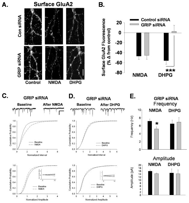
Figure 5. Knockdown of GRIP1/2 blocks mGluR-mediated AMPAR internalization and synaptic depression.
A. Surface GluA2 levels imaged after NMDA or DHPG treatment in neurons following knockdown of GRIP1/2 by siRNA. Neurons were transfected with scrambled or GRIP1/2 targeted siRNA and treated with NMDA or DHPG 72 hours later. While NMDAR-mediated reductions in surface GluA2 are unaffected by GRIP1/2 siRNA, the DHPG-induced decreases are blocked (Scale bar=10 μm). B. Graph shows quantitation of experiments represented in A. (n=8, ***P<0.001). C. The inter event interval (top) and amplitude (bottom) of mEPSCs was measured in GRIP siRNA transfected neurons before and 15 minutes after a 2 minute application of NMDA. As in control neurons, cumulative distribution plots show that NMDA caused an increase in intervals and decrease in amplitudes of mEPSCs in GRIP siRNA transfected neurons (n=5, Interval P<0.05; Amplitude P<0.01). Inset shows series resistance at 5 minutes before (−5) and 15 minutes (15) after agonist application. Scale bar for traces 500ms/20 pA. D. mEPSCs were recorded in cultured neurons during a 10 minute baseline period and 15 minutes after bath application of DHPG in GRIP siRNA transfected neurons. DHPG application caused no change in mEPSC inter event intervals (top) or amplitudes (bottom) in siRNA transfected neurons as indicated by the cumulative distribution plots. (n=5). E. Quantitation of median mEPSC frequency and amplitude experiments in C and D (*P<0.05).

