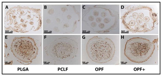
Figure 3.
NF-stained axons regenerated into the channels of scaffolds loaded with schwann cells 1 month after cord transection in rats.
Transverse section view of scaffold made from PLGA (A, 2/4 level), PCLF (B, 1/4 level), OPF (C, 3/4 level), and OPF+ (D, 3/4 level) polymers under microscope in lower magnification showing 7 channels. Transverse section view of scaffold made from PLGA (E, 1/4 level), PCLF (F, 1/4 level), OPF (G, 3/4 level), and OPF+ (H, 3/4 level) polymers under microscope in higher magnification showing a single channel.

