
Fig. 2.
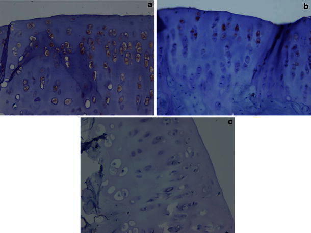
a Micrographs from group 1 (anterior cruciate ligament transection) distal femoral articular cartilage chondrocytes showing a high percentage of matrix metalloproteinase-3 (MMP-3) staining (MMP-3, immunoperoxidase ×20). b Micrographs from group 2 (anterior cruciate ligament transection + simvastatin) showing low percentage of MMP-3 staining (MMP-3, immunoperoxidase ×40). c Micrographs from group 3 (control knees) showing no MMP-3 staining (MMP-3, immunoperoxidase ×40)