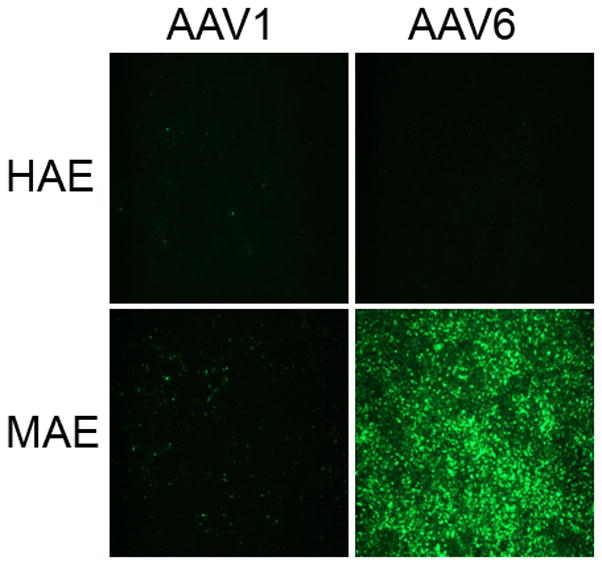
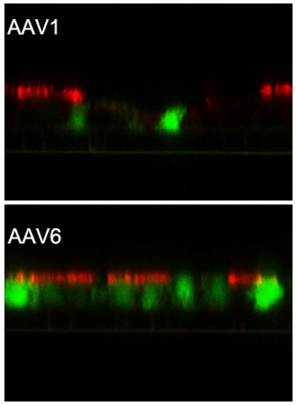
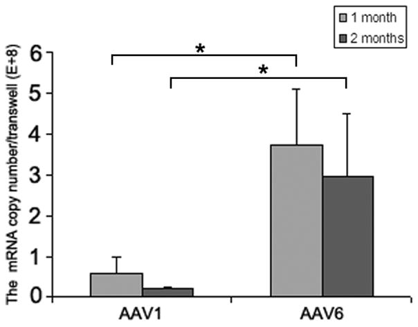
Fig. 1. Transduction efficiencies of AAV1 and AAV6 in human and mouse airway epithelial models in vitro.


Human and mouse airway epithelial cultures (HAE and MAE respectively) were inoculated apically with AAV1-GFP or AAV6-GFP at a dose of 1×10¹¹ vg/culture (1.0 cm2 surface area) (MOI = 1×10⁵). Representative en face fluorescence photomicrographs showing GFP-positive cells were obtained at 14 days post inoculation (p.i.). (a) Optical x-z confocal images of MAE transduced with AAV1-GFP and AAV6-GFP showing AAV-transduced cells as GFP positive. Cilia were immunolabeled with β-tubulin IV antibody (red) as described in “Material and Methods”. (b) The apical surface of MAE was inoculated with AAV1-GFP or AAV6-GFP (MOI of 5×10⁴). The mRNA from MAE was harvested at 1 and 2 months p.i., and relative GFP mRNA copy numbers were determined with qRT-PCR (as described in “Material and Methods”). Asterisk (*) denotes P<0.01. (c) AAV1-Luc or AAV6-Luc at a dose of 109 gc/culture (1 cm2) was applied to either the apical surface or the basolateral side of MAE and allowed to incubate at 37°C for 2 hrs. At 14 days p.i., luciferase activities were assessed, and presented as the mean (± SEM) luciferase activity per cm2 epithelia (n=4). RLU, relative light units. Asterisk (*) denotes P<0.01.
