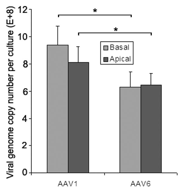
Fig. 4. AAV endocytosis at the apical and basolateral membranes.

AAV1 or AAV6 at MOI of 1×10⁴ was incubated with the apical or basolateral surfaces of MAE cultures at 37ºC for 2 hrs, followed by washing with DMEM three times to remove unbound virus. At 22 hrs p.i., Hirt DNA was extracted from infected cells and viral genome copies were assessed by quantitative PCR (qPCR). Data represent the mean ± SEM (n=4). Asterisk (*) denotes P<0.05.
