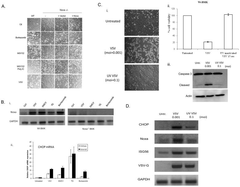
Fig. 3. Role of ER stress response in Noxa mode of action.
A) WT BMK cells, −/− cells, −/− cells transfected with empty vector and −/− cells stably expressing wild type Noxa were left untreated or treated with bortezomib (16 h, 20 nM), MG132 (6h, 10 μM), MG132 + poly IC (6h, 100 μg/ml) or infected with VSV (16 h, moi=0.001). After treatment, cells were photographed under phase contrast microscopy to illustrate morphological changes associated with those treatments. B) i. WT and −/− BMK cells were left untreated, infected with VSV (moi=0.001) or EMCV (moi=0.01) for 16 h, or treated with thapsigargin (TG; 1 μm) or bortezomib (20 nM) for 16h. RNA was isolated and RT-PCR studies were performed using primers specific for mouse Noxa and mouse GAPDH as a loading control. ii. CHOP (a marker of ER stress) mRNA levels were quantified by using real time SYBR green based qRT-PCR. C) i. WT BMK cells were left uninfected or were infected with replication competent VSV (moi=0.001) or UV-inactivated VSV (moi=0.1) for 16 h, after which time photomicrographs of the cells were taken under phase contrast microscopy. ii. Viability of cells treated as in “i” was determined by SRB staining, and results were presented as a percentage of the staining observed in uninfected controls. iii. Cells were infected as above and apoptosis demonstrated by immunoblot analysis of caspase 3 cleavage. D) Induction of ER stress-regulated and dsRNA-regulated genes was assessed by RT-PCR analysis of WT BMK cells infected with either wild type VSV (moi=0.001) or UV-inactivated virus (moi=0.1) 16 h after infection. Mouse GAPDH was used as an internal control.

