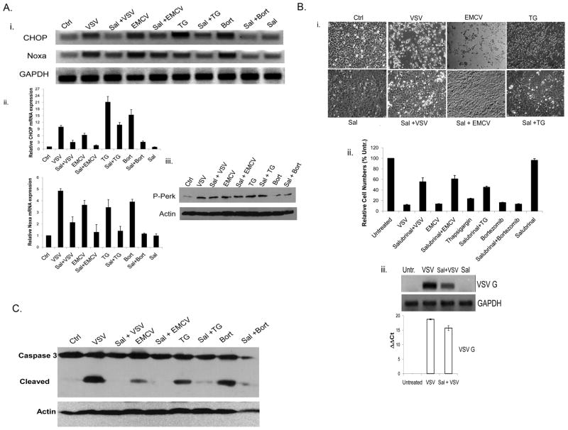
Fig. 4. Inhibition of ER stress response blocks Noxa upregulation and viral CPE.
A) i. WT BMK cells were left untreated (Ctrl) or were infected with VSV (16 h, moi=0.001), EMCV (16 h, moi=0.01), TG (16 h, 1 μM) or bortezomib (16 h, 20 nM) with or without prior treatment with the ER stress inhibitor salubrinal (Sal; 75 μM, 24 h). Total cellular RNA was then isolated and reverse transcribed for RT-PCR analysis of CHOP and Noxa mRNA induction. GAPDH was used as an internal control. ii. To quantify CHOP and Noxa mRNA expression, SYBR green-based qRT-PCR was used and the values were normalized to GAPDH. B) i. WT BMK cells were infected with VSV or EMCV or treated with TG, with or without Sal (75 μM) pretreatment. After 16 h, photomicrographs were taken to illustrate morphological changes associated with VSV, EMCV and TG cytopathicity.

