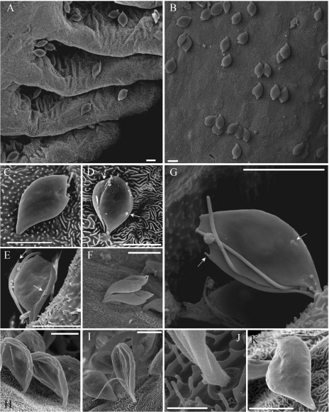
Fig. 4.
Scanning electron microscopy (SEM) of Ichthyobodo spp. trophozoites on gills of Atlantic salmon (Salmo salar). (A–G) Ichthyobodo salmonis sp. n. from brackishwater reared smolt. (A) Trophozoites attached to secondary gill lamellae; (B) trophozoites attached on the surface of a primary gill filament; (C) Single trophozoite, dorsal view; (D) trophozoite, ventral view; (E) quadri-flagellated (2 long, 2 short flagella) trophozoite, ventral view; (F) 2 trophozoites; (G) quadri-flagellated, ventral view (1short flagellum just visible in furrow); (H–K) Ichthyobodo necator s.s. from hatchery reared parr; (H) ventral view; (I) ventral view, flagella clearly visible; (J) same trophozoite shown in (I) close-up of the penetration area (scale bar=1 μm); (K) dorsal view; (D, E and G) arrows mark spine-like surface projections characteristic for I. salmonis sp.n. All scale bars except (J)=5 μm.

