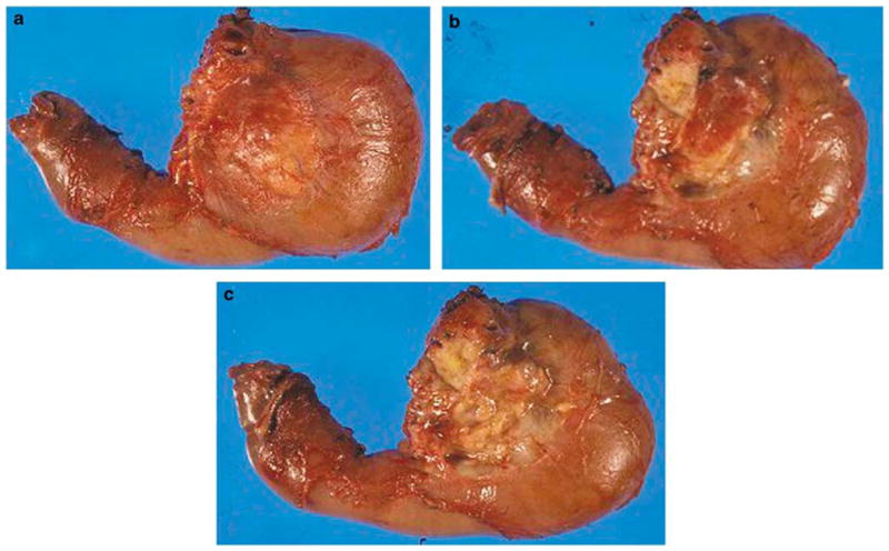
Figure 1.

Orange-peeling method. The peripancreatic soft tissues are dissected after margins were obtained but before sectioning of the pancreatic head. (a) Posterior surface of pancreatoduodenectomy specimen is seen before orange peeling. (b) The view after posterior pancreatoduodenal lymph node area (groove between pancreatic head and duodenal wall) is shaved off. (c) The view after posterior pancreatic surface is also orange peeled.
