
Figure 1.
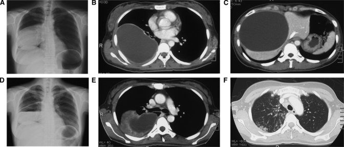
Chest radiography and abdominal tomography of this CE case. (A) A posterolateral chest radiograph revealed a well-circumscribed mass of 13 × 10 cm with homogenous density located on the right hemithorax. (B) On a CT scan of the thorax, a right lung lesion of 13 × 10 × 10 cm was noted. (C) Abdominal tomography showed a hepatic lesion with fluid density of 9 × 10 × 10 cm. (D) The wall of the lung cyst had started to degenerate and rupture at 14 days after the initiation of treatment. A water-fluid level was observed inside the cyst. (E and F) Eosinophilic infiltration appeared in both lung fields.