
Figure 2.
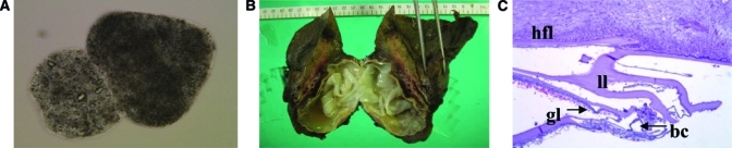
Parasitologic evidence of CE. (A) Protoscolices (0.2% trypan blue stain, 400×) in the aspirated fluid of both the lung and the hepatic cyst. (B) The lung CE cyst with a white outer membrane in the resected right lower lung lobe. (C) Microscopically, the cyst wall was composed of a brood capsule (bc), germinal layer (gl), laminated layer (ll), and hydatid fibrotic layer (hfl), (hematoxylin/eosin stain, 100x).