
Figure 7.
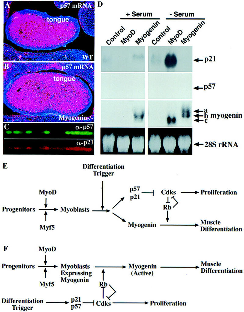
Myogenin is neither required nor sufficient for the expression of p21 and p57. (A,B) p57 expression detected with in situ hybridization on coronal sections of E15.5 embryos. (C) Colocalization of p21 and p57. Myotubes formed in vitro from primary embryonic myoblasts were stained with rabbit anti-p57 and goat anti-p21 polyclonal antibodies. p57 was visualized with a FITC-conjugated secondary antibody. p21 was visualized with a biotin-conjugated secondary antibody followed by Texas red-linked streptavidin. (D) A Northern blot of total RNA isolated from proliferating (+ serum) and differentiated (− serum) 10T1/2, MyoD-10T1/2, and myogenin–10T1/2 cells was probed sequentially with p21, p57, and myogenin. EtBr-stained 28S rRNA was used as a loading control. Three different sizes of myogenin mRNA are observed; a corresponds to the endogenous myogenin mRNA induced by myogenin, b corresponds to the myogenin transgene, and c corresponds to the endogenous myogenin mRNA induced by MyoD. The endogenous myogenin transcripts induced by MyoD and myogenin are of a different size. (E) A model for myogenesis in which myogenin, p21, and p57 are coordinately induced by a differentiation triggering signal to coordinate muscle cell differentiation. (F) A different model for myogenesis in which induction of p21 and p57 is the critical event that triggers muscle cell differentiation (see text for details).