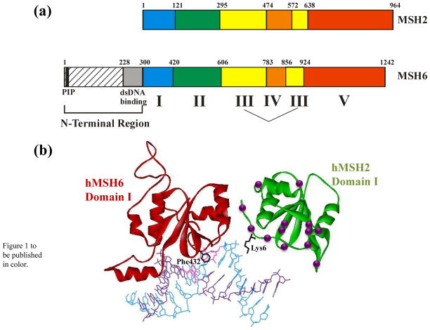
Figure 1.
a. Schematic of the domain structure of Msh2 and Msh6, based on homology with the human proteins.10 Domains I-V are defined by the MutS and MutSα crystal structures.10,22 The N-terminal Region is defined based on Clark et al.40 b. Structure of Domain I of hMSH2 (green) and hMH6 (red) with locations of HNPCC mutations in hMSH2 Domain I are indicated by the purple spheres. Lysine 6 of hMSH2 and the mispair-binding Phenylalanine 432 of hMSH6 are shown in black. The DNA mispair is colored pink. Figure was generated in WebLab Viewer using data from Warren et al.10

