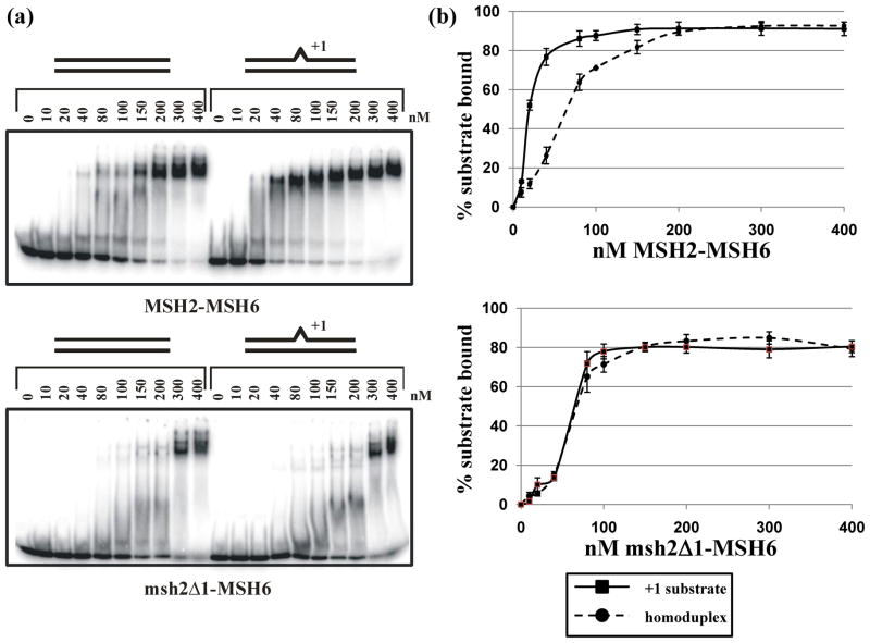
Figure 3.
Gel mobility shift assays of Msh2-Msh6 and msh2Δ1-Msh6. A. Titration of Msh2-Msh6 (top) and msh2Δ1-Msh6 (bottom) incubated with homoduplex and +1 substrates, as described in Materials and Methods. B. Quantification of gel mobility shift experiments, showing average binding of Msh2-Msh6 (top) and msh2Δ1-Msh6 (bottom) to a homoduplex or +1 substrate. The error bars indicate the standard error of the mean for four (Msh2-Msh6) or five (msh2Δ1-Msh6) independent experiments.

