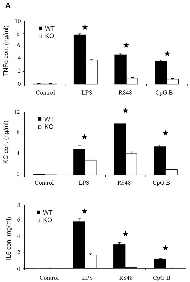
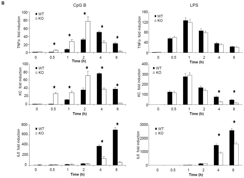
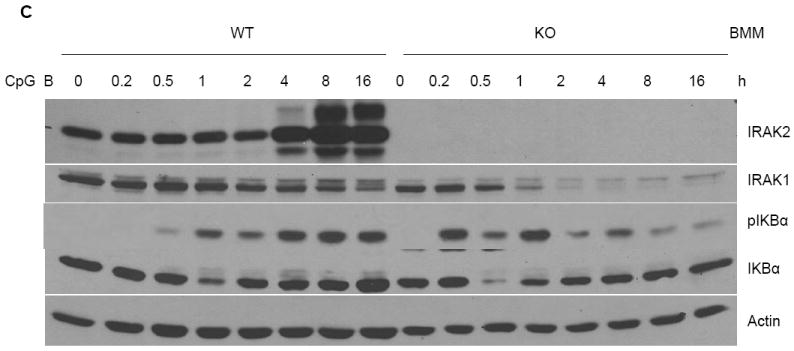
Figure 4. Impaired activation of NFκB at late times after CpG B stimulation in IRAK2-deficient macrophages.


(A) Wild-type and IRAK2-deficient BM-derived macrophages were treated with LPS (1 μg/ml), R848 (1 μg/ml), or CpG B (1 μg/ml) for 24 h. TNFα, KC and IL-6 concentrations in culture media were measured by ELISA. Results shown are the means and s. d. of triplicate determinations (* p< 0.05). (B) Wild-type and IRAK2-deficient BM-derived macrophages were treated with CpG B (1 μg/ml) or LPS (1 μg/ml) for the indicated times, and total RNAs (2 μg) were analyzed by real-time PCR to examine TNFα, KC and IL-6 expression (* p< 0.05). (C) Wild-type and IRAK2-deficient BM-derived macrophages were treated with CpG B (1 μg/ml) for the indicated times. Cell lysates were analyzed by the Western method with antibodies against IRAK2, IRAK1, phospho-IKBα, IKBα, and actin.
