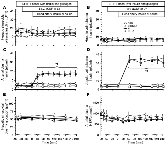
Figure 4. Plasma insulin, glucagon, and NEFA in dogs subjected to the head artery insulin infusion protocol.
As illustrated above graphs, 42-hour fasted conscious dogs received peripheral infusion of somatostatin and were maintained on a pancreatic clamp at basal levels of hepatic insulin, glucagon, and NEFA, then exposed to saline or insulin infusion into head arteries with i.c.v. infusion of either vehicle or LY. (A) Hepatic sinusoidal plasma insulin. (B) Hepatic vein plasma insulin. (C) Arterial plasma insulin. (D) Jugular vein plasma insulin. (E) Hepatic sinusoidal plasma glucagon. (F) Arterial plasma NEFA levels. Values are mean ± SEM. n = 4 (CTR and CTR+LY); 8 (HI); 7 (HI+LY). *P < 0.05, HI and HI+LY vs. CTR and CTR+LY; †P < 0.05, HI and HI+LY vs. basal period.

