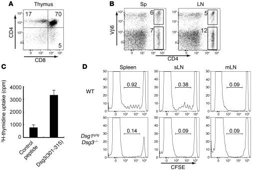
Figure 2. Generation of the Dsg3-specific TCR-transgenic mouse, Dsg3H1 mouse, and Dsg3 reactivity of transgenic T cells.
(A) Thymocytes were stained with anti-CD4 and -CD8 Abs and analyzed by flow cytometry. (B) Single-cell suspensions from the spleen and LNs were stained with anti-CD4 and -Vβ6 Abs and analyzed. (C) Splenocytes from Dsg3H1 mice were cultured with the peptide Dsg3(aa 301–315) or a control peptide. 3H-thymidine incorporation by these splenocytes is shown as the in vitro reactivity against Dsg3 peptide. (D) CFSE-labeled CD4+ T cells from Dsg3H1 mice were transferred into B6 WT mice and Dsg1tg/tgDsg3–/– mice. 3 days later, CFSE dilution was analyzed by flow cytometry after gating CD4+Vβ6+ cells of the spleen, skin-draining LN (sLN), and mesentery LN (mLN) from both recipients. Proportions of dividing cells were shown in each histogram. Similar results were obtained in 2 separate experiments. Data represent mean ± SEM.

