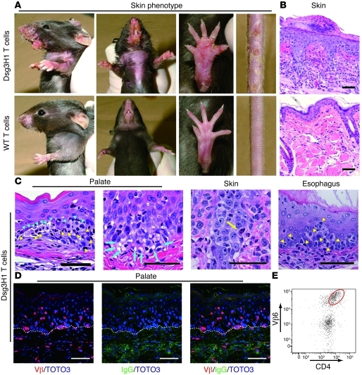
Figure 3. Tolerized Dsg3H1 T cells induced interface dermatitis, but not PV.
(A) The skin phenotype of Rag2–/– mice (n = 3 per group) given CD4+Vβ6+ cells from Dsg3H1 mice or CD4+ T cells from C57BL/6 mice in combination with Dsg3–/– B cells. Erosive and crusted lesion in the perioral region, upper limbs, paws, and tail and hair loss on the chest were evident (upper panels). (B) Pathology of the skin in the recipients given CD4+Vβ6+ cells from Dsg3H1 mice or WT CD4+ T cells. (C) Various Dsg3-expressing tissues in the recipients of CD4+Vβ6+ cells from Dsg3H1 mice were stained with H&E. Blue arrowheads indicate degenerated cells in the epithelium. Yellow arrowheads indicate intraepithelial inflammatory cells. Blue arrows indicate liquefaction degeneration in the basal layer of the lesional epithelium. Yellow arrow indicates a degenerated cell well stained with eosin, a so-called Civatte body. (D) The palate was stained with anti–TCR-β chain (red) and anti-IgG (green) Abs and TOTO3 (blue). Dotted lines indicate the basement membrane zone. (E) A single-cell suspension was prepared from the lesional skin of the recipients given CD4+Vβ6+ cells from Dsg3H1 mice and analyzed by flow cytometry after gating into the CD45+7-AAD– population. Scale bars: 50 μm.

