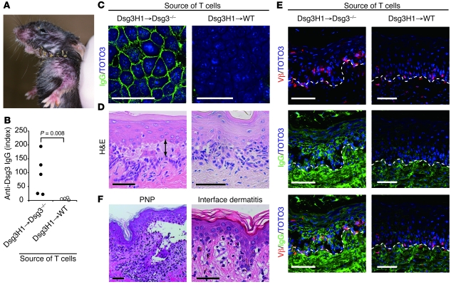
Figure 4. Nontolerized Dsg3H1 T cells induce both anti-Dsg3 IgG production and interface dermatitis.
BM cells from Dsg3H1 mice (Ly9.2) were transferred into sublethally irradiated 129/Sv WT and Dsg3–/– mice (Ly9.1) to generate BM chimeric mice, referred to as Dsg3H1→WT and Dsg3H1→Dsg3–/– mice, respectively. Donor-derived naive CD4+Vβ6+CD44–Ly9.1– cells were enriched from Dsg3H1→WT and Dsg3H1→Dsg3–/– mice and were cotransferred with Dsg3–/– B cells into Rag2–/– mice. (A) Skin phenotype in the recipient mice that received donor-derived CD4+ T cells from Dsg3H1→Dsg3–/– mice and Dsg3–/– B cells. Erythema and crusted lesions were observed in face, neck, and limbs. (B) Anti-Dsg3 IgG Ab titers in sera from the recipients were analyzed by ELISA. Statistical analysis was performed using the Mann-Whitney U test. (C) Sera from the recipients were added to the culture medium of Pam cells. IgG deposition on the cell surfaces was subsequently detected by anti-IgG Ab Alexa Fluor 488. Nuclei were stained with TOTO3 (blue). (D) Palates of the recipients were analyzed by H&E staining. The arrow indicates suprabasilar acantholysis. (E) Intraepidermal T cells (red) and IgG deposition (green) in the recipient’s palate were detected using anti–TCR-β Ab and anti-mouse IgG Ab, respectively. Dotted lines show the basement membrane zone. Similar results were obtained in 2 separate experiments. (F) The skin histopathology of PNP and interface dermatitis (GVHD) are shown. Scale bars: 50 μm.

