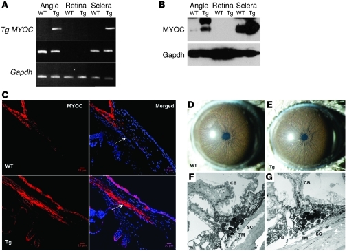
Figure 1. Increased transgene expression in TM of Tg-MYOCY437H mice.
Transgene (Y437H MYOC) and WT MYOC expression were examined in 6-month-old WT and Tg-MYOCY437H littermates by (A) RT-PCR, (B) Western blot analysis, and (C) immunostaining. PCR products were sequenced to confirm the presence of the transgene. Transgene and WT MYOC were expressed only in the iridocorneal angle and sclera of Tg-MYOCY437H mice but were absent in the retina of WT and Tg-MYOCY437H mice (n = 3). MYOC protein levels were increased in angle and sclera of Tg-MYOCY437H mice compared with WT littermates. Note that MYOC protein was absent in retinal lysates in Tg-MYOCY437H and WT mice (n = 3). GAPDH was used as a loading control. Immunostaining revealed that MYOC was localized to TM and is increased in Tg-MYOCY437H mice (bottom panels) compared with WT littermates (top panels). The TM is shown by the arrow. Scale bars: 20 μm. Open iridocorneal angle and normal morphology of anterior chamber structures in Tg-MYOCY437H mice (D–G). Slit lamp examination of anterior chambers of 6-month-old (D) WT mice and (E) Tg-MYOCY437H mice reveal no abnormalities in the iris, cornea, and lens. TEM images of the iridocorneal angle of 12-month-old (F) Tg-MYOCY437H mice were compared with (G) WT littermates. The iridocorneal angle is open in Tg-MYOCY437H mice (n = 6) compared with WT littermates (n = 5). Iris, ciliary body (CB), TM, and Schlemm canal (SC) are shown.

