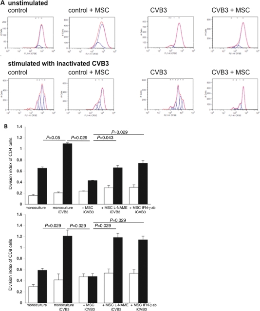
Figure 7.
Mesenchymal stem cells (MSCs) decrease cardiac mononuclear cell activation in coxsackievirus B3 (CVB3)-infected mice. (A) Cardiac mononuclear cells were isolated from control mice receiving PBS (Co + PBS) or MSCs (Co + mesenchymal stem cell), and of CVB3-infected mice receiving PBS (CVB3 + PBS) or mesenchymal stem cells (CVB3 + MSC). A minimum of n= 8 hearts/group were pooled to perform the experiment. Next, cardiac mononuclear cells were labelled with 10 µM of carboxyfluorescein succinimidyl ester to be able to measure cell proliferation and cultured for 72 h in the absence (unstimulated; upper panel) or presence of heat-inactivated CVB3 (stimulated: lower panel), followed by flow cytometry and analysis with FlowJo 8.7. software. Peaks are indicative for the amount of cell divisions. Representative peaks per group and condition ((un)stimulated) are shown. (B) Mononuclear cells were isolated from the spleen of control mice (open bar graph) or CVB3-infected (closed bar graph) mice. Next, carboxyfluorescein succinimidyl ester-labelled mononuclear cells were directly cultured, in the presence or absence of inactivated CVB3, with or without MSCs (untreated or 24 h pre-treated with L-NAME), in the presence or absence of 1 µg/mL of anti-murine interferon-γ (IFN-γ) antibody for 72 h. Then, cells were stained with monoclonal anti-CD4 or anti-CD8 antibodies, followed by flow cytometry and analysis with FlowJo 8.7. software. Bar graphs represent the division index of CD4− (upper panel) and of CD8− T cells (lower panel) with n= 4/group.

