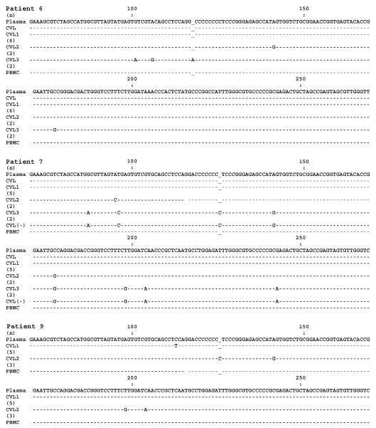
Figure 3.
Comparison of 5′ untranslated region hepatitis C virus (HCV) sequences amplified from plasma, cervicovaginal lavage (CVL) fluid, and peripheral-blood mononuclear cells (PBMCs) in patients 6, 7, and 9. In the figure, a minus sign indicates HCV RNA negative strand; “CVL” indicates the dominant variant; “CVL1,” “CVL2,” and “CVL3” indicate minor variants; and the nos. in parentheses indicate the no. of clones representing a given sequence.

