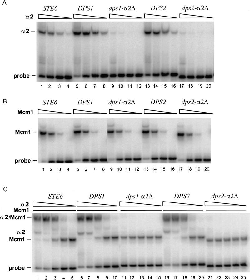
Figure 3.
DPS1 and DPS2 bind α2p and Mcm1p in vitro. (A) Binding of α2p to the indicated α2p/Mcm1p sites (sequences are shown in Table 1) as determined by EMSAs (described in Materials and Methods). Purified α2p was added to the binding reactions at 4.0 × 10⁻⁷ m (lanes 1,5,9,13,17), 8.0 × 10⁻⁸ m (lanes 2,6,10,14,18), 1.6 × m 10−8 m (lanes 3,7,11,15,19), or 3.2 × 10⁻⁹ m (lanes 4,8,12,16,20). (B) Binding of Mcm1p to the indicated site. Purified Mcm1p1-96 fragment was added to the binding reactions at 1.7 × 10⁻⁸ m (lanes 1,5,9,13,17), 1.7 × 10⁻⁹ m (lanes 2,6,10,14,18), 1.7 × 10⁻¹⁰ m (lanes 3,7,11,15,19), or 1.7 × 10⁻¹¹ m (lanes 4,8,12,16,20). (C) Binding of α2p and Mcm1p to the indicated site. Pruified α2p was added to the binding reactions at 8.0 × 10⁻⁸ m (lanes 1,6,11,16,21), 1.6 × 10⁻⁸ m (lanes 2,7,12,17,22), 3.2 × 10⁻⁹ m (lanes 3,8,13, 18,23), 6.4 × 10⁻¹⁰ m (lanes 4,9,14,19,24), or 1.3 × 10⁻¹⁰ m (lanes 5,10,15,20,25).

