Figure 5.
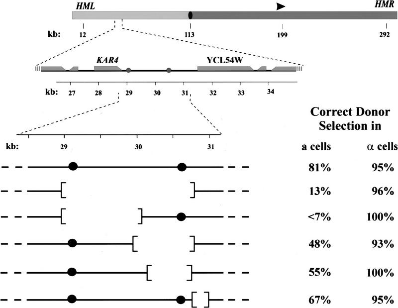
α2p/Mcm1p-binding sites on the left arm of chromosome III dictate donor preference in a cells. (Top) A diagram of chromosome III is shown which indicates the positions of HML, HMR, MAT (arrowhead), the centromere (filled oval), the left arm (light gray) and the right arm (dark gray). The positions of the three mating type loci and the centromere are specified (in kb from the left end of the chromosome). (Middle) The region from 26 to 35 kb of chromosome III is expanded to show the positions of the two α2p/Mcm1p-binding sites (shaded circles) and the adjacent open reading frames (shaded rectangles, taper at the 3′ end of the reading frame). (Bottom) The segment of chromosome III from 29.5 to 31 kb present in the wild-type (first line) and in several deletion strains, indicating the locations of the two α2p/Mcm1p sites (•) and the regions deleted in each strain. To the right of each chromosome segment is indicated the proportion of switching events in which the cell selected the appropriate locus (HML for a strains; HMR for α strains), determined as described in Materials and Methods.

