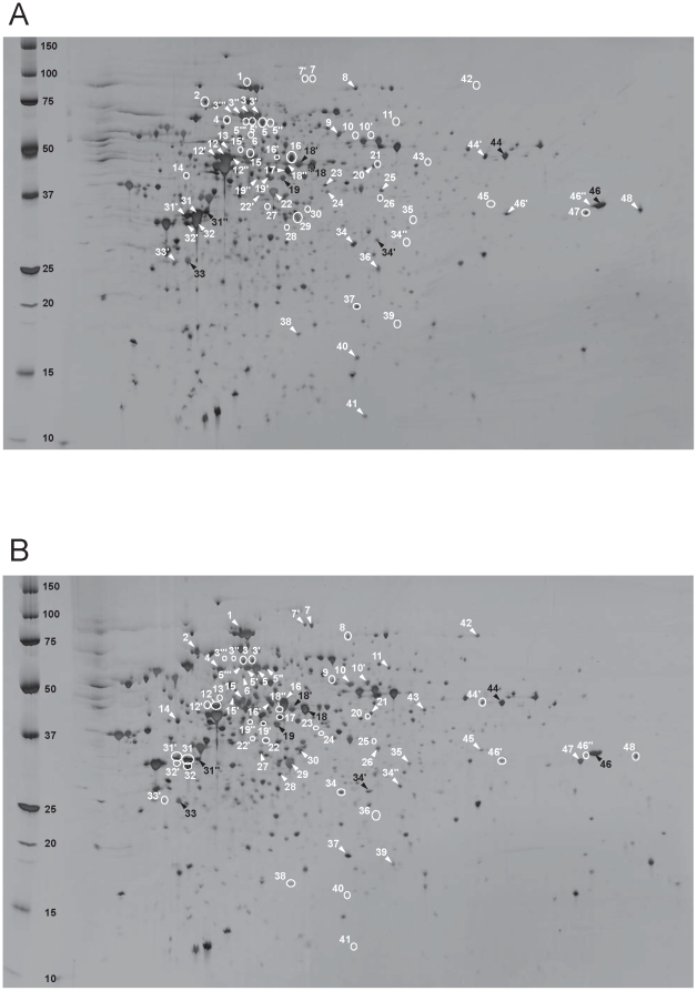
Figure 2. Comparative 2-DE analysis on pH range 4–7 of the proteins prepared from M. thermautotrophicus ΔH cells from (A) syntrophic co-culture and (B) pure-culture.
Crude protein extracts (40 mg) were separated on Immobiline dry strips pH 4–7 and SDS-PAGE on 12–14% polyacrylamide, and detected by silver staining. The peptide spots indicated by white arrows and white circles appeared with higher and lower intensity, respectively, than the corresponding peptide spots of the other culture condition. The black-numbered spots exhibited a similar expression level in both conditions.

