
Figure 1.
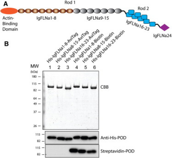
(A) Schematic structure of the FLNa monomer. The rod 1 segment contains domains IgFLNa 1–15 and the rod 2 segment contains domains IgFLNa 16–23. (B) SDS-PAGE gel of purified proteins used in this study. Proteins were stained with Coomassie brilliant blue (CBB) and detected by anti-His antibody and streptavidin conjugated with peroxidase (POD).