
Figure 3.
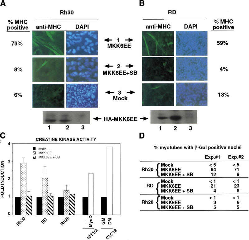


Introduction of MKK6EE reactivates myogenic differentiation in RMS cells. Two RMS cell lines, Rh30 (A) and RD (B), were infected with adenovirus expressing either the HA–MKK6EE gene or the β-gal gene (Mock) as described previously (Huang et al. 1997). Cells were transferred (12 hr postinfection) into DM with and without SB203580 (5 μm) for 96 hr and then fixed and stained for MHC or for DNA (DAPI). The percentage of MHC-positive multinucleated cells per field was counted and a representative value is reported. Expression of the exogenous MKK6EE proteins in the infected cells was shown by immunoblotting with anti-HA antibody. (C) Induction of CK activity in the indicated cell lines was assayed using a commercial kit (Sigma). (Solid bars) Mock; (stippled bars) MKK6EE; (hatched bars) MKK6EE + SB. The CK activity was calculated as micromoles of creatine formed/min per mg of protein extract. (D) RMS cells were first transfected with a reporter expressing β-gal fused to the nuclear localization signal (nls) and under the control of the MLC promoter (MLC–β-gal). After transfection (24 hr), the cells were infected with MKK6EE-expressing virus in the presence or absence of the p38 inhibitor SB. The percentage of myotubes with β-gal-positive nuclei is a measurement of multinucleation in myogenic cells. As a negative control cells were infected with an adenovirus expressing the β-gal gene (Mock) without the nls. Multinucleation was defined as a single cell with more than two β-gal-positive nuclei. Mock-infected cells were distinguished by the MKK6EE-infected cells as mononucleated cells with exclusively cytoplasmic blue staining. (E) The p21 luc reporter was transfected with the indicated plasmids in Rh30 (left) and RD (right) cells with or without SB (5 μm). At 18 hr post transfection, cells were transferred into DM and they were harvested after 24 hr. Luciferase activity was normalized to the expression levels of the transfected proteins (MyoD and MKK6EE were tagged with Flag and HA epitopes, respectively). The values shown are means and standard deviations from three independent experiments. (F) Plasmids expressing the indicated Gal4 fusion proteins were cotransfected with a gal4–luc reporter gene plus a plasmid expressing MKK6EE into RD and Rh30 cells growing in 20% FBS. After 12 hr, cells were placed in DM. MKK6EE cotransfected cells were also treated with 5 μm SB203580. Cells were harvested after 36 hr of culture in DM for LUC assay. The luciferase activity was normalized for the expression levels of the transfected Gal4 fusion proteins.