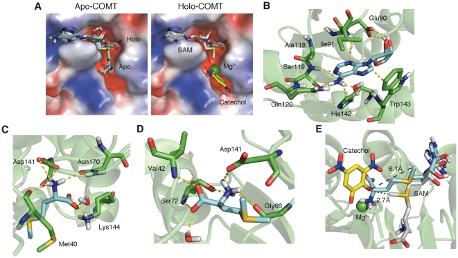
Figure 2. Binding configurations of SAM with and without metal.
(A) Alignment of SAM poses with and without metal. Cyan structure represents without metal (apo-COMT; derived from simulation) and gray structure represents SAM in the presence of metal (holo-COMT; from PDB ID 3BWM). In the right panel, the yellow structure is dinitrocatechol and green sphere is Zn2+. Surface of protein is shown as an electrostatic map with blue regions representing clusters of positive charge and red representing regions of negative charge. (B) Contacts made with adenosine portion of SAM (identical for apo- and holo-COMT). Cyan structure is adenosine and green structures are corresponding residues. Yellow dashed lines indicate contacts. (C) Contacts made with methionine portion of SAM (apo-COMT). Cyan structure is methionine portion of SAM. (D) Contacts made with methionine portion of SAM (holo-COMT). (E) Comparison of angle between methyl donor and accepting hydroxyl in the presence and absence of metal. Cyan structure is apo-COMT and gray structure is holo-COMT.

