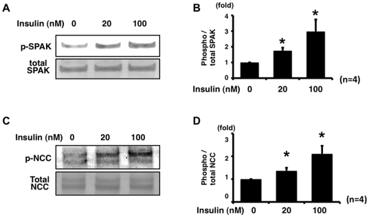
Figure 2. Insulin phosphorylates NCC and SPAK in mpkDCT cells in a dose-dependent manner.
A. Representative blot of phosphorylation of SPAK by insulin in mpkDCT cells. mpkDCT cells were incubated with insulin for 60 min at 20 nM and 100 nM. Insulin significantly increased phosphorylation of endogenous SPAK, compared to insulin-free control, in a dose-dependent manner. B. Densitometry analysis of phosphorylation of SPAK by insulin in mpkDCT cells. Values (n = 4) are expressed as the ratio to the signals in insulin-free samples. *P<0.05. C. Representative blot of phosphorylation of NCC by insulin in mpkDCT cells. Insulin significantly increased phosphorylation of endogenous NCC, compared to insulin-free control, in a dose-dependent manner. D. Densitometry analysis of phosphorylation of NCC by insulin in mpkDCT cells. Values (n = 4) are expressed as the ratio to the signals in insulin-free samples. *P<0.05.

