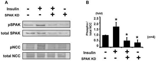
Figure 3. Insulin-induced phosphorylation of NCC was impaired in SPAK knockdown mpkDCT cells.
A. Representative blot of phosphorylation of NCC by insulin in SPAK knockdown mpkDCT cells. mpkDCT cells were incubated with insulin for 60 min at 100 nM. Insulin-induced NCC phosphorylation was impaired in SPAK knockdown mpkDCT cells, although insulin increased phosphorylation of NCC in negative control siRNA-transfected cells. B. Densitometry analysis of phosphorylation of NCC by insulin in negative control and SPAK knockdown mpkDCT cells. Values (n = 4) are expressed as the ratio to the signals in insulin-free negative control siRNA-transfected cells.

