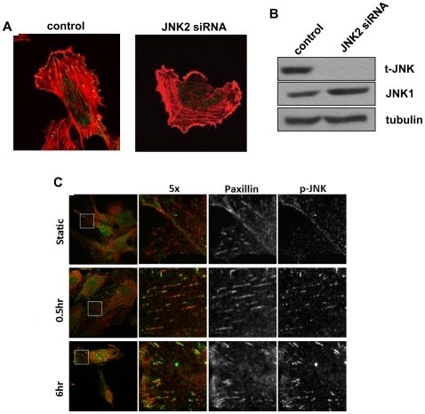
Figure 3. Activated JNK in focal adhesions.
(A) Specific staining for activated JNK. HUVECs transfected with either control or JNK2 siRNA for 48 h were plated on FN-coated coverslips 2 hours, then fixed and stained for phospho-JNK (green) and actin (red). (B) Western blot of JNK2 knockdown. (C) HUVECs were left untreated or exposed to laminar shear stress as indicated. Cells were stained for phospho-JNK (green) and paxillin (red). Results are representative of 3 experiments.

