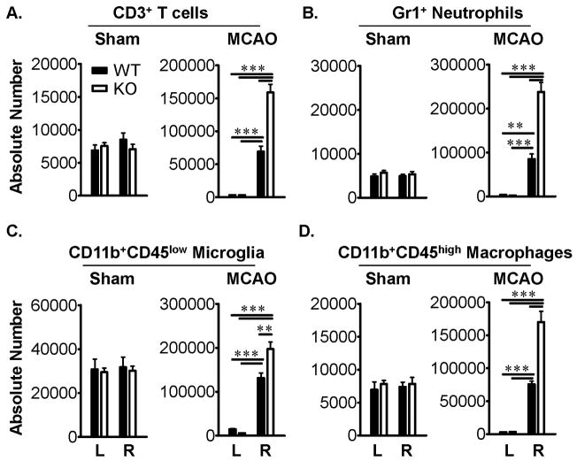
Figure 3. PD-1 reduces the infiltration of inflammatory cells into the ischemic brain after MCAO.
Absolute numbers of CD3+ T cells (A), Gr1+ neutrophils (B), CD11b+CD45low microglia (C), and CD11b+CD45high macrophages (D) were significantly increased (**, P<0.001; ***, P<0.0001) in ipsilateral right (R) but not contralateral left (L) brain hemispheres of PD-1-KO versus WT mice after 60min MCAO and 96h reperfusion but not Sham-treatment. Values represent Mean ± SEM from five mice per group.

