Figure 1.
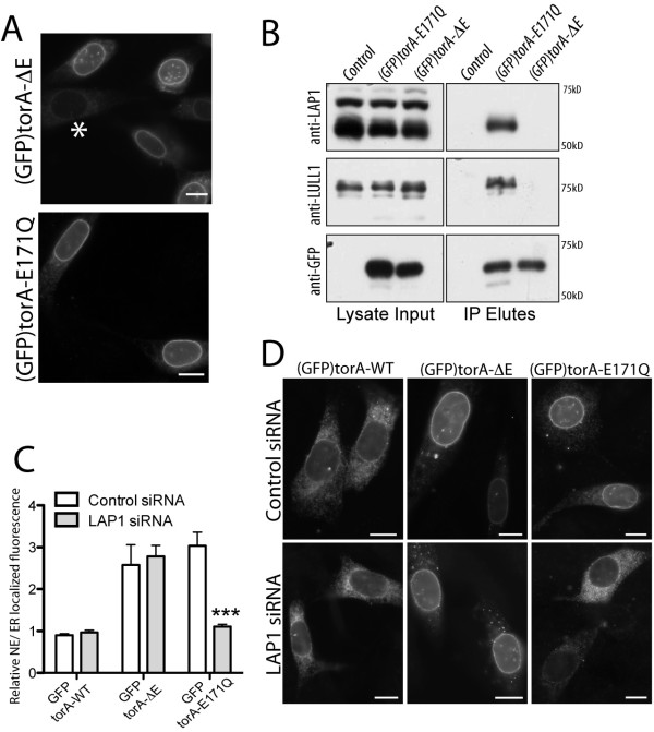
TorA-ΔE does not interact with the NE-localized torA-E171Q binding partner, LAP1. (A) (GFP)torA-ΔE and (GFP)torA-E171Q both concentrate in the NE of NIH-3T3 cells. The * symbol highlights a cell with ER-localized (GFP)torA-ΔE. Scale bars show 10 μm. (B) LAP1 and LULL1 co-immunoprecipitate with (GFP)torA-E171Q and not (GFP)torA-ΔE. Left panels show levels of LAP1, LULL1 and (GFP)torA in 5% of lysate input collected from control, (GFP)torA-E171Q or (GFP)torA-ΔE stably expressing NIH-3T3 cells. Anti-LAP1 (top row) detects the alternatively spliced LAP1 isoforms [14,43] and anti-LULL1 (middle row) detects 75 kD LULL1. Right panels show levels of LAP1, LULL1, or GFP that immunoprecipitate with anti-GFP in buffer containing 2 mM ATP and 0.5 mM ADP. Note: mild elution conditions were used to prevent release of captured anti-GFP IgG molecules. (C) LAP1 depletion specifically affects (GFP)torA-E171Q localization. Columns show the mean and s.e.m. for ratios of NE/ER localized GFP fluorescence from n > 25 NIH-3T3 cells co-transfected with scrambled or LAP1 siRNA duplexes and (GFP)torA isoforms. *** symbol indicates that LAP1 depletion significantly reduces the (GFP)torA-E171Q NE/ER fluorescence ratio (p < 0.001; Two-Way ANOVA with Bonferroni post-hoc analysis). (D) (GFP)torA-ΔE remains NE-localized in LAP1 depleted cells, while (GFP)torA-E171Q redistributes into the ER. Images show anti-GFP labeling of NIH-3T3 cells co-transfected with (GFP)torA isoforms and control (upper row) or LAP1 siRNA duplexes (lower row). Scale bars show 10 μm.

