
Figure 7.
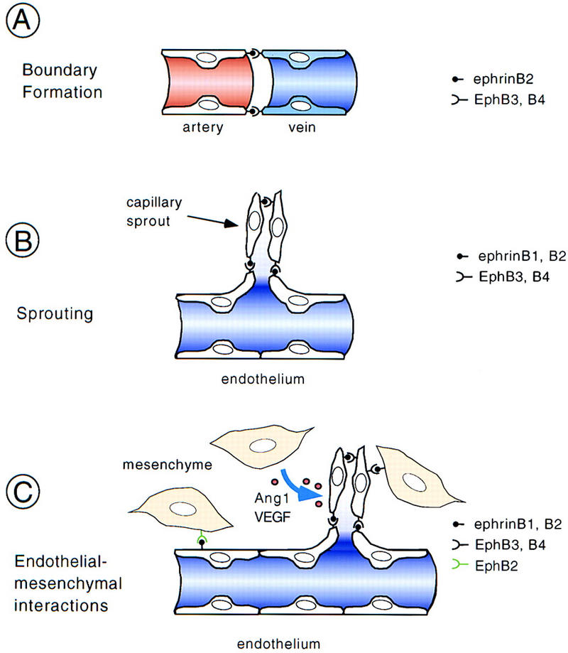
Presumed mechanisms and sites of action of ephrins and Eph receptors during remodeling of the vasculature. (A) Interaction of ephrinB2 ligand expressed on arteries and EphB3 and EphB4 receptors expressed on veins demarcates the boundary between arterial and venous domains. By analogy to the action in the nervous system, it was suggested that ephrin–Eph interactions may prevent intermixing of arterial and venous endothelial cells and, following sprouting, may result in the formation of a capillary network (Yancopoulos et al. 1998). However, this model was based on the exclusive and complementary expression of ephrinB2 and EphB4. (B) Coexpression of ligands and receptors on the same type of vessels (e.g., veins) provides a cell-to-cell signal for endothelial cells that may rather be stimulatory and help to promote morphogenesis and sprouting. (C) Mesenchymal cells adjacent to blood vessels also express ephrins or Eph receptors and may help patterning the vasculature. In the somites, this signal may be inhibitory and prevent sprouting, whereas in other regions, stimulatory signals are conceivable. Mesenchymal cells are also the source of angiogenic factors such as Ang1 and VEGF, which may modulate ephrinB–EphB receptor signaling.