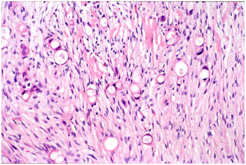
Fig. 4.

In areas where the infiltrative units are smaller, tangential cut of the vacuolated glands may give the impression of individual cell infiltration mimicking diffuse infiltrative-type (“poorly cohesive cell” or “signet-ring” cell) carcinoma; however, the presence of larger cohesive glandular elements establish the diagnosis that this is merely the vacuolated variant of ductal adenocarcinoma
