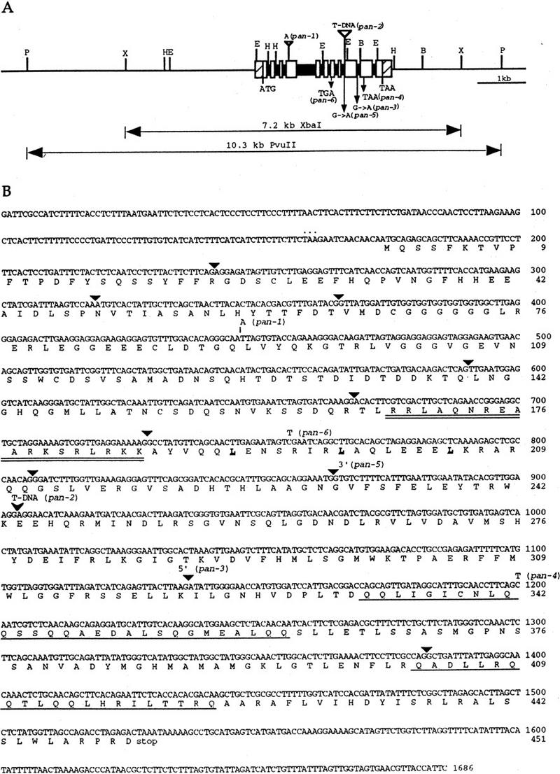
Figure 2.

Genomic structure of the PAN gene, and sequence of the PAN cDNA. (A) Structure and restriction map of the PAN genomic region. The open boxes and solid boxes represent exons and introns, respectively. The hatched boxes represent the 5′ and 3′ untranslated regions. The genomic regions used for complementation of pan mutant phenotype are indicated below. ATG and TAA indicate the positions of the putative translational start and stop codons, respectively. Mutations in the six pan alleles are also shown. Restriction sites are indicated as follows: (B) BamHI; (E) EcoRI; (H) HindIII; (P) PvuII; (X) XbaI. (B) The cDNA and deduced amino acid sequence of the PAN gene. The predicted amino acid sequence for the longest open reading frame is shown directly below the nucleotide sequence. Numbers to the right of the sequence refer to the positions of nucleotide and amino acid residues. Triangles indicate the positions of introns. The basic region is doubly underlined and the leucines in the leucine zipper are in bold and underlined. The glutamine-rich regions are singly underlined. The inframe stop codon preceding the first methionine is indicated by dots above the nucleotide sequence. One nucleotide insertion in pan-1 is shown above the nucleotide sequence. The position of the T-DNA insertion in pan-2 allele is indicated by an arrowhead. The pan-3 mutation occurs at the splice donor site of the ninth intron. The pan-5 allele is mutated in the splice acceptor site of the eighth intron. Point mutations resulting in premature translational stop sites in pan-4 and pan-6 are shown above the nucleotide sequence.
