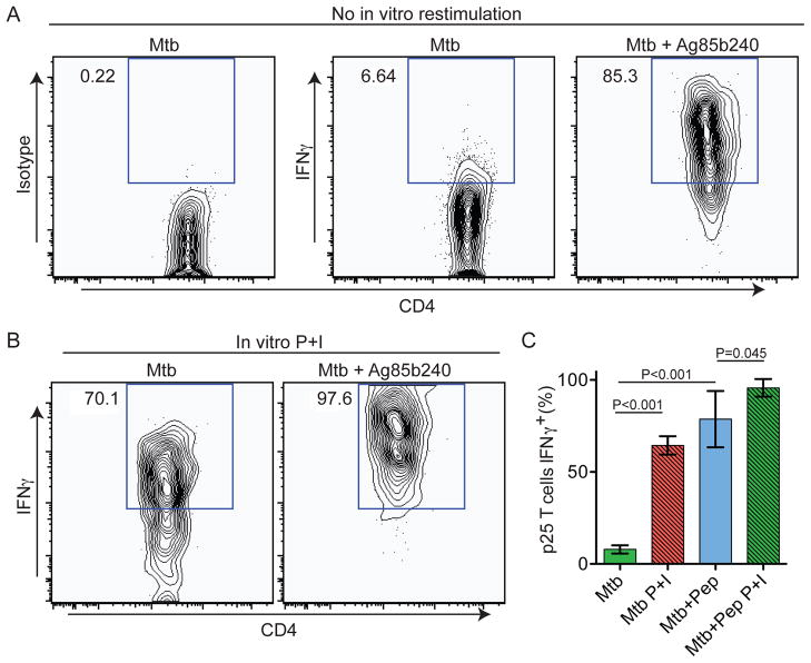
Figure 7. Antigen-specific T cells show limited effector function but high effector potential in Mtb-induced granulomas.
(A) Animals were treated as in Figure 4 except they were infected with Mtb. Representative histograms gated on CD4+EGFP+ cells from p25-EGFP-bearing infected animals that had been injected with PBS or Ag85b peptide 2 hours prior to analysis. (B) IHLs were restimulated with P+I prior to analysis in order to detect maximum cytokine production. (C) Quantification of percentage of p25 T cells expressing IFN-γ. Graph shows mean+/− SD from at least three mice per group and is representative of two similar experiments. P values from a two-tailed T test are shown.

