Fig. 4.
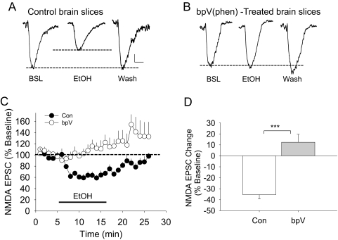
Inhibition of protein tyrosine phosphatases can block ethanol inhibition of the NMDAR currents. Brain slices were treated with 10 μM bpV(phen) for 3 h and were tested for the effects of a short-term 80 mM ethanol challenge. Representative traces show that ethanol (80 mM, EtOH) inhibits NMDA EPSCs in control brain slices (A) compared with bpV(phen)-treated brain slices (B). Dotted lines represent baseline and the extent of ethanol inhibition. BSL, baseline; Wash, washout. C, time course of the average responses for synaptic NMDA EPSC amplitudes from control (Con) (n = 8) or bpV-treated brain slice preparations (n = 8). D, bar graphs show the averaged effects of 80 mM ethanol on NMDA EPSCs from control (Con, − 35.4 ± 3.9%) or from bpV(phen)-treated (12.4 ± 7.3%) slices. There is a significant difference between these two groups (t = 5.808, p < 0.001, Student's t test). Scale bars, 20 pA and 25 ms. ***, p < 0.001.

