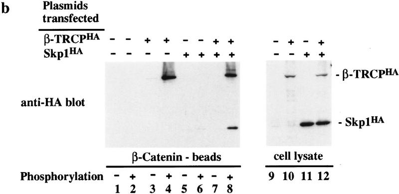
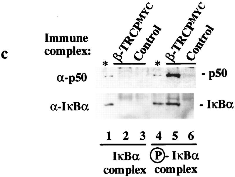
Figure 3.
Association of SCFβ-TRCP with IκBα and β-catenin destruction motifs and with the IκBα/NF-κB complex. (a,b) Cell lysates (0.3 μg of protein/150 μl) from Fig. 2 were used in IκBα (a) and β-catenin (b) peptide bead binding reactions as described in Materials and Methods. Bound proteins were analyzed by immunoblotting with the indicated antibodies. (c) Phosphorylation-dependent association of β-TRCPMyc with the IκBα/p50/p65 complex in vitro. β-TRCPMyc immune complexes (lanes 2,5) corresponding to those in Fig. 2a (lane 3) or control complexes (lanes 3,6) corresponding to those in Fig. 2a (lane 1) were used in binding reactions with either IκBα/p50/p65 or IκK-β phosphorylated IκBα/p50/p65 complexes (see Materials and Methods). Bound proteins were separated by SDS-PAGE and immunoblotted using anti-p50 or anti-IκBα antibodies. The asterisk (lanes 1,4) indicates the positions of 15% of the input IκBα complexes used in the binding reaction.


