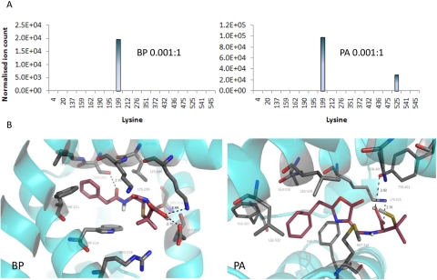
Fig. 4.
Selective binding of BP or PA to HSA identified in vitro. A, at low concentration BP preferentially bound to Lys199, whereas PA bound to Lys199 and Lys525. B, molecular modeling of noncovalent interaction of drug with HSA revealed the best poses by docking BP and PA into HSA, showing the key proximity between Lys199 and the β-lactam carbonyl group for BP, and Lys525 and the oxazolone ring for PA. Proteins are rendered as cyan ribbons, amino acid residues close to the guest molecule are rendered as sticks (carbon, gray; nitrogen, blue; oxygen, red), and guest molecules are rendered as sticks (carbon, violet; nitrogen, blue; oxygen, red; polar hydrogens, white).

