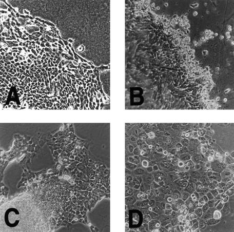
Figure 1.
Morphology of TCC compared to HUC in culture. (A) Normal HUCs show typical epithelial morphology with tightly adherent monolayers of polygonal cell. Note the lack of mesenchymal cell contamination. Original magnification, 200×. (B) Superficial papillary TCCs also show similar epithelial cellular morphology in vitro, but unlike normal HUCs, often form three dimensional structures at the leading edge of growth reminiscent of their in vivo multilayered morphology. Original magnification, 200×. (C) Myoinvasive papillary TCC 96-2 also formed a cell line that retained its ability to form three-dimensional structures after prolonged culture. Original magnification, 200×. (D) Myoinvasive TCC 96-1 with flat morphology in vitro is shown here. Original magnification, 200×.

