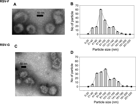
Figure 2.
Viruslike particles (VLPs) containing M1 and respiratory syncytial virus (RSV) F (A) or RSV G (C) visualized and their size distribution determined (B, D) after negative staining EM (H-7500, Hitachi). Fifty-five (55%) of RSV F VLPs from 170 particles were distributed between 60 and 100 nm (B), and 66% of RSV-G VLPs from 161 particles were distributed between the size 40 and 100 nm (D).

