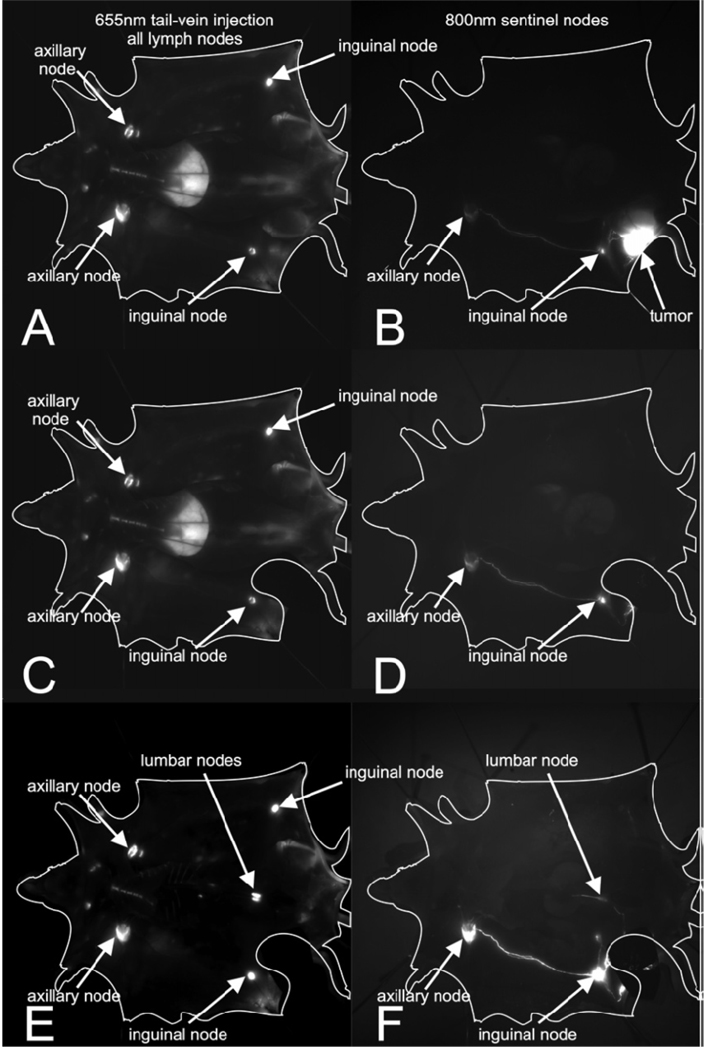
Fig 2.
Fluorescent imaging lymphatic drainage after injection of fluorescent QDs into mice bearing M21 melanoma. Left frames A,C,E: channel after tail vein injection of PEG 5k-COOH coated QDs; right frames B,DE: 800-nm channel after intratumoral injection of PEG 5k-OMe coated QDs.[35]

