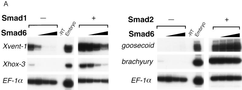
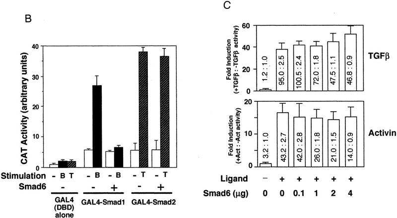
Figure 3.
Smad6 specifically inhibits the BMP/Smad1 signaling pathway. (A) Smad6 blocks induction of ventral mesoderm by Smad1 but not induction of dorsal mesoderm by Smad2. An increasing amount of Smad6 RNA (0.5–4 ng) was coinjected with or without a fixed amount of Smad1 or Smad2 RNA (2 ng). Animal caps from injected embryos were collected at gastrula stage (stage 11.5) and subjected to RT–PCR. Xvent-1 and Xhox-3 are markers of ventral mesoderm; goosecoid is a dorsal mesoderm marker and brachyury is a pan–mesodermal marker. (B) BMP-dependent transcriptional activation of GAL4–Smad1 fusion protein is blocked by Smad6. R-1B/L17 cells were transfected with the reporter gene (G1E1BCAT, 1 μg), appropriate receptors (TβR-I for TGFβ or BMPR–IB and BMPR–II for BMP2), and either a vector containing the GAL4 DNA-binding domain (DBD) alone or fusion constructs of DBD with Smad1 or Smad2 in the presence or absence of Smad6 (2 μg). Cells were incubated with or without 5 nm BMP2 (B) or 100 pm TGFβ (T) for 18 hr. CAT activity is expressed as the mean ± s.d. of three independent experiments. (C) TGFβ or activin-induced transcriptional activation of the 3TP promoter is not inhibited by Smad6. R-1B/L17 cells were transfected with the reporter gene (p3TP–lux) and type I receptors for TGFβ (TβR-l) or activin (ActR-IB) and were treated with or without 100 pm TGFβ (top) or 2 nm activin (bottom) for 18 hr. The ratios of stimulated to unstimulated levels of luciferase activity are indicated numerically, and their quotients plotted in the bar graph. Data are the mean ± s.d. of triplicate values. Notice that although Smad6 transfection decreased the basal as well as the agonist-induced levels of luciferase activity, it did not decrease the relative induction by TGFβ or activin.


