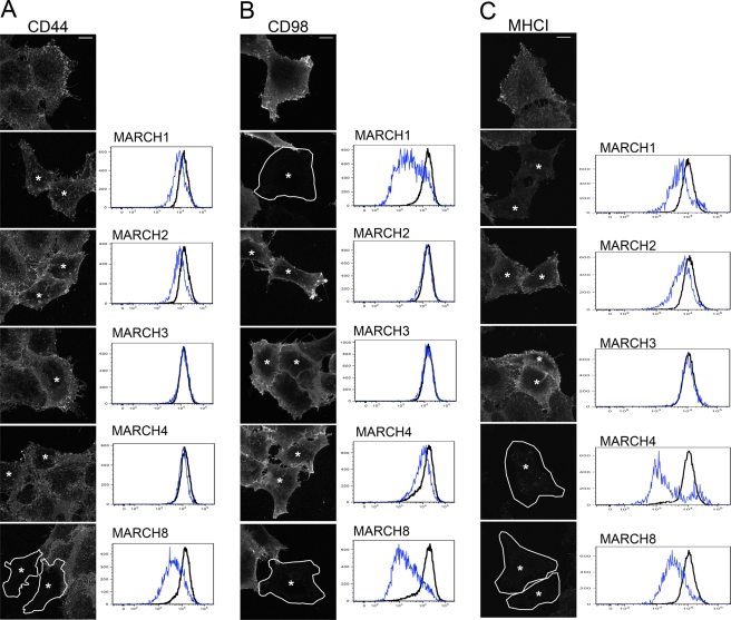
FIGURE 3:
Effect of MARCH protein expression on surface levels of CD44, CD98, and MHCI. HeLa cells were transfected with the indicated MARCH-FLAG construct as described. After 18 h, the surface of the cells was labeled with mouse antibodies to CD44, CD98, and MHCI and processed for either immunofluorescence or flow cytometry. In fluorescence images, MARCH-transfected cells (as detected by rabbit anti-FLAG staining; not shown) are indicated by an asterisk and outlined in some cases. Graphs show the Alexa 488 staining intensity (x-axis) versus cell number (y-axis) for surface CD44 (A), CD98 (B), or MHCI (C) in control cells (black line) vs. indicated MARCH-expressing cells (blue line) from flow cytometry analysis. Bars, 10 μm.

