Fig. 7.
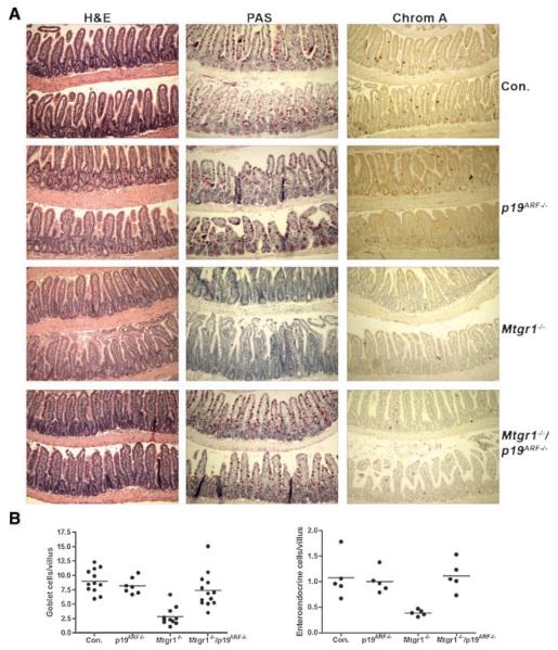
Loss of p19ARF restores the secretory lineage in mice lacking Mtgr1. A: H&E stained sections of ilea from adult wild-type (Con.), Mtgr1−/−, p19ARF−/−, and Mtgr1−/−/p19ARF−/− mice (100× magnification). PAS staining of the ilea of adult mice to detect Goblet cells, which stain fuchsia (100× magnification) and immunohistochemical staining with anti-Chromogranin A to detect enteroendocrine cells in dark brown (100× magnification). A larger view of the Mtgr1−/− and double-null panels for PAS can be seen in Supplemental Figure 1. B: Quantification of the results in (A) shown in graphical form. The numbers of-positive cells were counted over at least 100 villi per mouse with mean value indicated with a solid line. For PAS, control mice were compared to Mtgr1−/− mice, P = 0.0001; for Mtgr1−/− compared to Mtgr1−/−/p19ARF−/−, P = 0.0002. For Chromagranin A, comparison of control vs. Mtgr1−/−, P = 0.0067; Mtgr1−/− vs. Mtgr1−/−/p19ARF−/− P = 0.0007.

